During Muscle Contraction Myosin Crossbridges Bind To Active Sites On
Kalali
Mar 13, 2025 · 7 min read
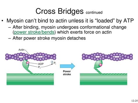
Table of Contents
During Muscle Contraction: Myosin Crossbridges Bind to Active Sites on Actin
The human body is a marvel of biological engineering, capable of a wide range of movements, from the delicate tap of a finger to the powerful stride of a runner. This remarkable capacity is largely due to the intricate workings of our muscles, microscopic machines that convert chemical energy into mechanical work. At the heart of muscle contraction lies a complex interplay between proteins, primarily actin and myosin, whose interaction forms the basis of muscle function. This article delves deep into the fascinating process of muscle contraction, focusing specifically on the crucial role of myosin crossbridges binding to active sites on actin.
Understanding the Players: Actin and Myosin
Before we explore the binding process, let's briefly introduce the key players. Muscle fibers are composed of repeating units called sarcomeres, the fundamental units of muscle contraction. Within the sarcomere, two primary proteins orchestrate the contraction process:
Actin: The Thin Filament
Actin filaments are thin, helical structures that form a significant part of the sarcomere's structure. These filaments are not simply linear; they are studded with myosin-binding sites, also known as active sites. These sites are crucial because they are the points of interaction with myosin. Crucially, these active sites are usually blocked by tropomyosin, a protein that prevents continuous muscle contraction.
Myosin: The Thick Filament
Myosin filaments are thicker and have a distinctive structure. Each myosin molecule is a dimer, with two globular heads (myosin heads or crossbridges) and a long tail. These globular heads possess ATPase activity, meaning they can hydrolyze adenosine triphosphate (ATP) – the body's energy currency – to power muscle contraction. The myosin heads are the crucial components that interact with the active sites on actin.
The Sliding Filament Theory: A Foundation for Understanding
The sliding filament theory explains the mechanism of muscle contraction. It proposes that muscle contraction occurs through the sliding of thin (actin) filaments over thick (myosin) filaments, resulting in a shortening of the sarcomere and overall muscle fiber. This sliding is precisely controlled by the cyclical interaction of myosin crossbridges with actin's active sites.
The Cycle of Crossbridge Binding and Muscle Contraction: A Detailed Look
The process of muscle contraction is a dynamic cycle involving several distinct steps:
1. ATP Hydrolysis and Crossbridge Activation:
The cycle begins with a myosin head bound to ADP and inorganic phosphate (Pi). The myosin head is in a high-energy conformation, "cocked" and ready to bind. The binding of a new ATP molecule to the myosin head causes a conformational change, releasing ADP and Pi. This conformational change is crucial; it causes the myosin head to detach from the actin active site.
2. Crossbridge Binding:
Now, in its high-energy state, the myosin head searches for a nearby actin active site. The presence of calcium ions (Ca²⁺) plays a crucial role here. Ca²⁺ binds to troponin, another protein associated with actin and tropomyosin. This binding causes a conformational shift in tropomyosin, exposing the myosin-binding sites on actin. The myosin head can now bind to an available active site on the actin filament.
3. Power Stroke:
Once the myosin head binds to the active site, the release of Pi triggers the power stroke. This is a conformational change in the myosin head, causing it to pivot and pull the actin filament towards the center of the sarcomere. This sliding movement is the fundamental mechanism behind muscle contraction. The myosin head is now in a low-energy state.
4. ATP Binding and Detachment:
Following the power stroke, the myosin head remains bound to the actin filament in its low-energy state. A new ATP molecule binds to the myosin head, causing it to detach from the actin active site. This detachment is essential for the cycle to repeat.
5. Cycle Repetition:
The cycle of ATP hydrolysis, crossbridge binding, the power stroke, and detachment repeats numerous times, with countless myosin heads simultaneously engaging in this process. Each cycle generates a small movement, but the cumulative effect of thousands of cycles results in significant muscle shortening and force generation.
The Role of Calcium Ions (Ca²⁺) and Regulation of Muscle Contraction
The precise regulation of muscle contraction is paramount. It's not a simple on/off switch but a highly controlled process. Calcium ions (Ca²⁺) are the key regulators of this intricate system. When a motor neuron stimulates a muscle fiber, it triggers the release of Ca²⁺ from the sarcoplasmic reticulum (SR), a specialized intracellular calcium store.
The rise in cytosolic Ca²⁺ concentration initiates the events described above. As mentioned, Ca²⁺ binds to troponin, causing a conformational change in tropomyosin that uncovers the myosin-binding sites on actin. This allows myosin crossbridges to interact with actin and initiate the cycle of muscle contraction.
When the neural signal ceases, Ca²⁺ is actively pumped back into the SR, causing tropomyosin to return to its blocking position. This prevents further interaction between myosin and actin, effectively terminating the muscle contraction.
The Importance of ATP in Muscle Contraction
ATP is the energy source powering the entire cycle. It plays a pivotal role in three crucial stages:
- Myosin Head Detachment: ATP binding is essential for the detachment of the myosin head from the actin active site, allowing the cycle to continue.
- Crossbridge Activation: ATP hydrolysis provides the energy for the conformational change in the myosin head, leading to the high-energy state ready for binding.
- Calcium Pump Activity: ATP fuels the active transport of Ca²⁺ back into the SR, relaxing the muscle.
Without sufficient ATP, the myosin heads would remain bound to the actin filaments, resulting in a state of rigor – a condition known as rigor mortis in deceased individuals.
Types of Muscle Contraction: Isometric and Isotonic
Muscle contractions can be broadly classified into two types:
Isometric Contractions:
In isometric contractions, muscle length remains constant, but muscle tension increases. This type of contraction is seen when holding an object still, where the force generated by the muscle is counteracted by an external force, preventing movement. The crossbridge cycle still occurs, but the overall muscle length does not change.
Isotonic Contractions:
In isotonic contractions, muscle tension remains relatively constant, but muscle length changes. This type of contraction is responsible for most movements, such as lifting a weight or walking. The crossbridge cycle is highly active, leading to either shortening (concentric contraction) or lengthening (eccentric contraction) of the muscle.
Muscle Fatigue and its Relationship to Crossbridge Cycling
Muscle fatigue, the decline in muscle force or power output during sustained or repetitive activity, is a complex phenomenon involving several factors. While the precise mechanisms are still being investigated, it is clear that the crossbridge cycle is impacted. Fatigue is believed to result from various factors including depletion of energy reserves (ATP), accumulation of metabolic by-products (lactate), and disturbances in ion homeostasis (Ca²⁺, K⁺). These factors can reduce the efficiency of the crossbridge cycle, impairing the ability of the muscle to generate force.
Diseases and Disorders Affecting Muscle Contraction
Several diseases and disorders can impair the process of muscle contraction. These include:
-
Muscular Dystrophy: A group of genetic disorders characterized by progressive muscle weakness and degeneration. These conditions often affect the structural proteins within the muscle, disrupting the integrity of the sarcomere and the ability of myosin crossbridges to effectively interact with actin.
-
Myasthenia Gravis: An autoimmune disorder affecting the neuromuscular junction, the site where motor neurons communicate with muscle fibers. Antibodies interfere with the transmission of signals from the neuron to the muscle, reducing the ability of the muscle to contract effectively.
-
Amyotrophic Lateral Sclerosis (ALS): A progressive neurodegenerative disease affecting motor neurons, leading to muscle weakness and atrophy. The loss of motor neuron function impairs the signaling required for proper muscle contraction.
Conclusion: The Intricate Dance of Actin and Myosin
The binding of myosin crossbridges to active sites on actin is a fundamental process underlying muscle contraction. This finely-tuned mechanism, regulated by calcium ions and driven by ATP, allows for the generation of force and movement. A thorough understanding of this process is vital for comprehending not only the mechanics of movement but also the pathogenesis of various muscle disorders. Future research in this area promises to shed further light on this intricate biological dance and pave the way for more effective treatments and therapies for muscle-related diseases.
Latest Posts
Latest Posts
-
How Old Am I If Born In 1986
Jul 06, 2025
-
Where Is Food Coloring In The Grocery Store
Jul 06, 2025
-
How Do You Say Pickles In Spanish
Jul 06, 2025
-
How Much Is 1000 Hours In Days
Jul 06, 2025
-
Sic A Parrot On The Guild Emissary
Jul 06, 2025
Related Post
Thank you for visiting our website which covers about During Muscle Contraction Myosin Crossbridges Bind To Active Sites On . We hope the information provided has been useful to you. Feel free to contact us if you have any questions or need further assistance. See you next time and don't miss to bookmark.