In Fully Contracted Muscles The Actin Filaments Lie Side-by-side.
Kalali
Mar 19, 2025 · 6 min read
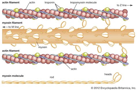
Table of Contents
In Fully Contracted Muscles, the Actin Filaments Lie Side-by-Side: A Deep Dive into Muscle Contraction
The intricate dance of muscle contraction, a fundamental process enabling movement and life itself, involves a complex interplay of proteins and structural changes. A key aspect often overlooked in simplified explanations is the final arrangement of the actin filaments within a fully contracted muscle fiber. Contrary to some initial impressions, in a fully contracted state, the actin filaments do not simply overlap extensively; instead, they lie side-by-side, a crucial detail that profoundly impacts our understanding of the limits of muscle shortening. This article will delve deeply into the mechanism of muscle contraction, focusing on the final, often-missed, arrangement of actin filaments and the implications thereof.
Understanding the Sliding Filament Theory
Before exploring the side-by-side arrangement, we must first solidify our understanding of the sliding filament theory, the cornerstone of muscle contraction mechanics. This theory postulates that muscle contraction results from the relative sliding of actin and myosin filaments over each other, without any change in the length of the individual filaments themselves.
The Key Players: Actin and Myosin
-
Actin Filaments: These thin filaments are composed primarily of actin monomers, arranged in a double helix structure. Tropomyosin and troponin are crucial regulatory proteins associated with actin, controlling the interaction with myosin.
-
Myosin Filaments: These thick filaments are composed of numerous myosin molecules, each with a head and tail region. The myosin heads possess ATPase activity, essential for generating the force of contraction.
The Cross-Bridge Cycle: A Detailed Look
The sliding filament theory is fueled by the cyclical interaction between actin and myosin, known as the cross-bridge cycle. This involves several key steps:
-
Attachment: The myosin head, in its high-energy conformation (ADP and Pi bound), binds to an actin molecule, forming a cross-bridge.
-
Power Stroke: The release of Pi triggers a conformational change in the myosin head, causing it to swivel and pull the actin filament towards the center of the sarcomere (the basic contractile unit of muscle).
-
Detachment: The binding of ATP to the myosin head weakens the actin-myosin bond, causing detachment.
-
Reactivation: ATP hydrolysis resets the myosin head to its high-energy conformation, preparing it for another cycle.
This continuous cycle of attachment, power stroke, detachment, and reactivation, across numerous cross-bridges within a sarcomere, drives the sliding of actin and myosin filaments, resulting in muscle shortening and force generation.
The Significance of Sarcomere Structure
Understanding the sarcomere's structural organization is vital for comprehending the final arrangement of actin filaments in a fully contracted muscle. The sarcomere is defined by its boundaries, the Z-lines, to which actin filaments are anchored. Myosin filaments are located centrally, overlapping with the actin filaments.
The H-Zone and I-Band: Indicators of Contraction
Two key structural components, the H-zone and the I-band, provide visual cues about the degree of muscle contraction:
-
H-zone: The region in the center of the sarcomere occupied solely by myosin filaments. It decreases in size during contraction as actin filaments slide inward.
-
I-band: The region containing only actin filaments. It also shrinks during contraction as actin filaments overlap more extensively with myosin filaments.
In a fully relaxed muscle, both the H-zone and I-band are relatively wide. As contraction progresses, these zones narrow until, in a fully contracted state, the H-zone virtually disappears, and the I-band is significantly reduced. This narrowing isn't simply due to overlapping actin filaments; it's a crucial prelude to the side-by-side arrangement.
The Side-by-Side Arrangement: The Final Stage
While the significant overlap of actin and myosin filaments is evident in moderately contracted muscles, reaching the fully contracted state reveals a fascinating arrangement. The actin filaments, having slid as far as structurally possible toward the center of the sarcomere, ultimately come to rest side-by-side.
This side-by-side arrangement is a consequence of several factors:
-
Steric Hindrance: The physical limitations imposed by the thick myosin filaments prevent further interdigitation of actin filaments. The myosin filaments act as a barrier, forcing the actin filaments to align alongside each other.
-
Z-line Structure: The structural integrity of the Z-lines, anchoring points for the actin filaments, prevents further inward movement beyond a certain point.
-
Titin's Role: The giant protein titin, which runs along the length of the sarcomere, acts as a molecular spring and plays a role in defining the limits of sarcomere shortening. Its elasticity contributes to preventing excessive overlap and encourages side-by-side arrangement.
This side-by-side arrangement isn't simply an accidental consequence; it is a critical aspect of the muscle's biomechanical properties. It dictates the maximum shortening capacity of the muscle fiber and plays a role in force generation at the limits of contraction.
Implications of the Side-by-Side Arrangement
The final side-by-side arrangement of actin filaments in a fully contracted muscle has several important implications:
-
Maximum Shortening: This arrangement dictates the maximum degree to which a muscle fiber can shorten. It defines the physiological limit of muscle contraction.
-
Force Generation: While the force generated may plateau or even slightly decrease at the fully contracted state due to reduced cross-bridge formation opportunities, the side-by-side arrangement ensures that the force generated is still significant and provides structural stability.
-
Muscle Stiffness: The tightly packed arrangement of actin filaments in the fully contracted state contributes to the increased stiffness of the muscle.
Understanding this side-by-side arrangement is crucial for fields such as biomechanics, physiology, and medicine. It provides insights into:
-
Muscle Injury: Forces exceeding the capacity of the muscle to shorten and maintain the side-by-side arrangement can lead to injury.
-
Muscle Disease: Conditions affecting the structure and function of actin, myosin, or other sarcomeric proteins can alter the dynamics of contraction and the final arrangement of filaments.
-
Artificial Muscle Design: Knowledge of the structural limits and the side-by-side arrangement informs the design of artificial muscles and bio-inspired materials.
Conclusion: Beyond Simple Overlap
The commonly simplified depiction of muscle contraction as solely an overlap of actin and myosin filaments is incomplete. The reality is far more nuanced, culminating in the fascinating side-by-side arrangement of actin filaments in fully contracted muscles. This arrangement is not merely a final state but a critical element shaping the biomechanics, physiology, and ultimately, the function of muscle tissue. By appreciating this detail, we gain a deeper understanding of the intricate dance of muscle contraction and its limitations, opening avenues for further research and innovation in related fields. Further research continues to unravel the intricacies of this process, focusing on the roles of individual proteins, the effects of various stimuli, and the subtle variations in arrangement across different muscle types. The quest to fully understand muscle contraction is far from over, and the side-by-side arrangement remains a key piece in this ongoing scientific puzzle.
Latest Posts
Latest Posts
-
Twenty Is What Percent Of 200
Mar 20, 2025
-
How Do You Write A Polynomial In Standard Form
Mar 20, 2025
-
How Many Pints Is In A Litre
Mar 20, 2025
-
How Many Feet Is 17 Inches
Mar 20, 2025
-
2 To The Power Of 4
Mar 20, 2025
Related Post
Thank you for visiting our website which covers about In Fully Contracted Muscles The Actin Filaments Lie Side-by-side. . We hope the information provided has been useful to you. Feel free to contact us if you have any questions or need further assistance. See you next time and don't miss to bookmark.
