Receptors For Hearing Are Located In The
Kalali
Mar 27, 2025 · 6 min read
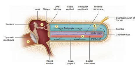
Table of Contents
Receptors for Hearing are Located in the Cochlea: A Deep Dive into Auditory Perception
Hearing, a fundamental sense, allows us to perceive the world around us through sound. This incredible ability relies on a complex interplay of structures within the ear, culminating in the intricate workings of the cochlea. This article delves deep into the fascinating world of auditory perception, focusing specifically on the location and function of the hair cells, the crucial receptors for hearing, nestled within the cochlea.
The Journey of Sound: From Outer Ear to Cochlea
Before we explore the cochlea's inner workings, let's briefly trace the path of sound waves as they travel through the ear:
1. The Outer Ear: Collection and Funneling
The journey begins in the outer ear, comprising the pinna (the visible part of the ear) and the external auditory canal. The pinna acts like a satellite dish, collecting sound waves and funneling them into the canal towards the eardrum.
2. The Middle Ear: Amplification and Transmission
The sound waves reach the tympanic membrane (eardrum), causing it to vibrate. These vibrations are then amplified by the three tiny bones of the middle ear—the malleus (hammer), incus (anvil), and stapes (stirrup)—acting as a lever system. This amplification is crucial for transmitting sound effectively to the inner ear. The stapes, the innermost bone, transmits the vibrations to the oval window, a membrane separating the middle and inner ear.
3. The Inner Ear: Transduction into Neural Signals
The inner ear, a fluid-filled labyrinth, houses the cochlea, a snail-shaped structure where the magic of auditory transduction happens. The vibrations from the stapes create pressure waves in the fluid within the cochlea, setting the stage for the activation of the hair cells.
The Cochlea: The Organ of Hearing
The cochlea is a fascinating structure, crucial for our ability to hear. Its intricate anatomy enables it to discriminate between different frequencies of sound. Let's break down its key components:
1. The Spiral Structure and Fluid-Filled Chambers
The cochlea is coiled like a snail shell, with approximately 2.5 turns. Internally, it's divided into three fluid-filled chambers:
- Scala vestibuli: The upper chamber, connected to the oval window.
- Scala media (cochlear duct): The middle chamber, containing the organ of Corti, home to the hair cells. It's filled with endolymph, a fluid with a high potassium concentration.
- Scala tympani: The lower chamber, connected to the round window. Like the scala vestibuli, it's filled with perilymph, a fluid similar to cerebrospinal fluid.
2. The Basilar Membrane: Frequency Discrimination
The basilar membrane is a crucial structure within the scala media, separating it from the scala tympani. This membrane is not uniform; it's wider and more flexible at its apex (the furthest point from the oval window) and narrower and stiffer at its base (closest to the oval window). This difference in stiffness allows for tonotopic organization: high-frequency sounds cause maximal vibration near the base, while low-frequency sounds cause maximal vibration near the apex. This is fundamental to our ability to distinguish between different pitches.
3. The Organ of Corti: The Sensory Epithelium
Sitting atop the basilar membrane is the organ of Corti, the sensory epithelium of hearing. This structure houses the hair cells, the actual receptors for hearing. It's a remarkably complex arrangement of cells and supporting structures, ensuring efficient transduction of mechanical vibrations into electrical signals.
Hair Cells: The Transducers of Sound
The hair cells are the sensory receptors responsible for converting mechanical vibrations into electrical signals that the brain can interpret as sound. There are two main types:
1. Inner Hair Cells (IHCs): Primary Transducers
Inner hair cells (IHCs) are the primary transducers of sound. They are arranged in a single row along the length of the basilar membrane. Their stereocilia (hair-like structures) are arranged in a precise “V” formation, crucial for their function. When the basilar membrane vibrates, the stereocilia bend, opening mechanically gated ion channels. This influx of ions causes depolarization of the hair cell, leading to the release of neurotransmitters onto the auditory nerve fibers. These fibers transmit the electrical signals to the brainstem, initiating the process of auditory perception.
2. Outer Hair Cells (OHCs): Amplification and Fine-Tuning
Outer hair cells (OHCs) are arranged in three rows along the basilar membrane. Unlike IHCs, OHCs play a more active role in auditory processing. They possess a unique motor protein, prestin, which allows them to change their length in response to changes in membrane potential. This electromotility amplifies the movement of the basilar membrane, enhancing the sensitivity and frequency selectivity of the cochlea. This amplification is particularly important for detecting low-intensity sounds. OHCs also contribute to the sharpness of our hearing, enhancing our ability to distinguish between similar frequencies.
The Auditory Nerve: Transmission to the Brain
The signals generated by the hair cells are transmitted to the brain via the auditory nerve, a bundle of nerve fibers that originates from the cochlea. The auditory nerve fibers synapse with the hair cells, and their firing patterns reflect the frequency and intensity of the sound. This information is then relayed through a series of nuclei in the brainstem, midbrain, and thalamus, ultimately reaching the auditory cortex in the temporal lobe, where sound is perceived and interpreted.
Age-Related Hearing Loss and Hair Cell Damage
Age-related hearing loss, or presbycusis, is a common condition affecting many older adults. One of the major contributors to presbycusis is the progressive loss of hair cells, particularly outer hair cells. This loss reduces the amplification and frequency selectivity of the cochlea, resulting in impaired hearing, especially for high-frequency sounds. The exact mechanisms underlying age-related hair cell loss are not fully understood, but factors such as oxidative stress, inflammation, and genetic predisposition likely play a significant role.
Noise-Induced Hearing Loss and Hair Cell Damage
Exposure to loud noises can also cause significant damage to hair cells, leading to noise-induced hearing loss (NIHL). Loud sounds can overwhelm the cochlea, causing mechanical damage to hair cells and their supporting structures. This damage can be temporary (temporary threshold shift) or permanent (permanent threshold shift), depending on the intensity and duration of exposure. Prolonged exposure to loud noises can lead to progressive hearing loss, tinnitus (ringing in the ears), and hyperacusis (increased sensitivity to sound).
Protecting Your Hearing
Protecting your hearing is crucial for maintaining your quality of life. Here are some key steps you can take:
- Reduce exposure to loud noises: Use earplugs or earmuffs in noisy environments, such as concerts, construction sites, and factories.
- Turn down the volume: Keep the volume of your headphones and other audio devices at a safe level.
- Get regular hearing checkups: Early detection of hearing loss can help prevent further damage and improve treatment outcomes.
- Be aware of the risks of certain medications: Some medications can have ototoxic (hearing-damaging) side effects. Discuss any concerns with your doctor.
Conclusion: The Intricate Symphony of Sound
The receptors for hearing are located within the intricate structure of the cochlea, specifically within the organ of Corti. The hair cells, both inner and outer, are the key players in converting mechanical vibrations into electrical signals, allowing us to perceive the rich tapestry of sounds in our world. Understanding the anatomy and physiology of the cochlea and the role of hair cells is crucial for appreciating the complexity of auditory perception and for developing strategies to prevent and treat hearing loss. The delicate balance within the cochlea, and the remarkable sensitivity of hair cells, highlights the intricate marvel of our auditory system. Preserving the health of these crucial receptors is vital for maintaining the precious gift of hearing.
Latest Posts
Latest Posts
-
8 Pints Is How Many Cups
Mar 30, 2025
-
30 Meters Is How Many Feet
Mar 30, 2025
-
What Is 30 Percent Of 800
Mar 30, 2025
-
How Many Liters Is 100 Ml
Mar 30, 2025
-
1 In 25 As A Percentage
Mar 30, 2025
Related Post
Thank you for visiting our website which covers about Receptors For Hearing Are Located In The . We hope the information provided has been useful to you. Feel free to contact us if you have any questions or need further assistance. See you next time and don't miss to bookmark.
