The Shaft Of A Long Bone Is Known As The
Kalali
Mar 11, 2025 · 6 min read
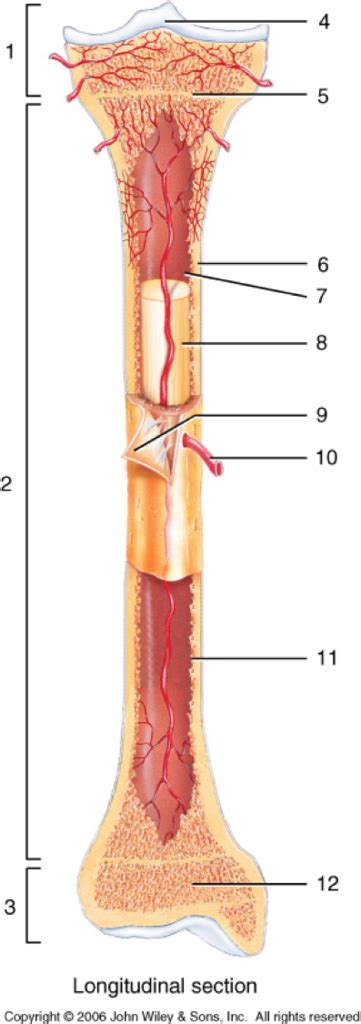
Table of Contents
The Shaft of a Long Bone is Known as the Diaphysis: A Deep Dive into Long Bone Anatomy
The human skeletal system, a marvel of biological engineering, provides structure, support, and protection for our bodies. Understanding its intricacies is crucial for anyone interested in anatomy, physiology, or related fields. A key component of this system is the long bone, and within the long bone lies a crucial structural element: the diaphysis. This article will delve deep into the diaphysis, exploring its structure, function, and significance in overall skeletal health.
What is a Diaphysis?
The diaphysis, also known as the shaft, is the long, cylindrical main portion of a long bone. It's the central part of the bone, located between the two ends. Think of it as the main body of the bone, providing structural integrity and acting as a lever arm for muscle attachment. Unlike the ends of the bone (epiphyses), the diaphysis is primarily composed of compact bone, providing maximum strength and rigidity. This compact bone tissue is dense and solid, enabling the bone to withstand significant forces.
Differentiating Diaphysis from Epiphysis and Metaphysis
To fully grasp the diaphysis's role, it's essential to differentiate it from other parts of the long bone:
-
Epiphysis: This is the expanded end of a long bone. The epiphyses contain spongy bone, which is less dense than compact bone but is still crucial for strength and weight distribution. They are covered with articular cartilage, which allows for smooth joint movement. The epiphyses are responsible for much of the bone's growth in length during childhood and adolescence.
-
Metaphysis: This is the transitional region between the diaphysis and epiphysis. It contains the epiphyseal plate (growth plate) in growing bones. This plate is responsible for the longitudinal growth of the bone. Once growth is complete, the epiphyseal plate closes, leaving behind the epiphyseal line.
The clear distinction between these three parts—diaphysis, metaphysis, and epiphysis—is critical for understanding bone growth, development, and potential injury sites.
The Microscopic Structure of the Diaphysis: Compact Bone Tissue
The diaphysis's strength and resilience stem from its predominantly compact bone composition. Let's examine the microscopic structure to appreciate its robust nature:
-
Osteons (Haversian Systems): These are the fundamental functional units of compact bone. Each osteon is a cylindrical structure composed of concentric lamellae (rings) of bone tissue surrounding a central Haversian canal. These canals contain blood vessels and nerves that supply the bone cells.
-
Lamellae: These are thin layers of bone matrix, organized in concentric circles around the Haversian canals. They provide strength and support to the bone. Interstitial lamellae are remnants of old osteons, while circumferential lamellae encircle the entire diaphysis.
-
Lacunae: These are small spaces within the bone matrix where osteocytes (bone cells) reside. Osteocytes are responsible for maintaining the bone tissue.
-
Canaliculi: These are tiny canals that connect the lacunae, allowing osteocytes to communicate and exchange nutrients and waste products.
This intricate arrangement of osteons, lamellae, lacunae, and canaliculi creates a highly organized and incredibly strong structure within the diaphysis, capable of withstanding immense stress and pressure.
The Diaphysis's Crucial Role in Locomotion and Support
The diaphysis plays a critical role in several vital functions:
-
Weight Bearing: The diaphysis acts as the primary weight-bearing component of long bones. Its strong, compact structure can withstand the forces generated by body weight and movement.
-
Leverage: The long, cylindrical shape of the diaphysis acts as a lever arm for muscle action. Muscles attach to the bone via tendons, and the diaphysis allows for efficient transmission of forces during movement. This leverage is essential for locomotion, manipulation of objects, and maintaining posture.
-
Protection of the Medulla: The diaphysis protects the bone marrow, located within the medullary cavity. This cavity is a hollow space within the diaphysis that houses red bone marrow (responsible for blood cell production) in younger individuals and yellow bone marrow (primarily fat storage) in adults.
-
Mineral Storage: Bone tissue throughout the diaphysis acts as a reservoir for essential minerals, such as calcium and phosphorus. These minerals are crucial for various physiological processes, including muscle contraction, nerve transmission, and blood clotting. The body can draw upon these reserves when necessary.
Common Injuries and Conditions Affecting the Diaphysis
The diaphysis, while robust, is still susceptible to injury and certain conditions:
-
Fractures: Diaphyseal fractures are common, particularly in long bones like the femur, tibia, and humerus. These fractures can result from trauma, high-impact activities, or underlying bone diseases. The severity of a diaphyseal fracture can vary, ranging from simple cracks to complete breaks.
-
Stress Fractures: These are tiny cracks in the bone caused by repetitive stress, such as overuse in athletic activities. They often occur in the diaphysis and can be painful and debilitating.
-
Bone Infections (Osteomyelitis): Infection of the bone tissue, which can affect the diaphysis, often requiring aggressive treatment.
-
Bone Tumors: Primary or secondary bone tumors can occur in the diaphysis, requiring diagnosis and appropriate management.
-
Bone Density Loss (Osteoporosis): This condition weakens the bones, making the diaphysis more vulnerable to fractures.
Diaphyseal Development and Growth
The development of the diaphysis is a complex process that involves several stages:
-
Intramembranous Ossification: Certain bones, while not strictly long bones, form directly from mesenchymal tissue through intramembranous ossification. While not directly relevant to the diaphysis of long bones, understanding this process provides a wider context to bone development.
-
Endochondral Ossification: This is the primary process involved in long bone formation, including the diaphysis. It involves the replacement of a cartilaginous model with bone tissue. The diaphysis is the first part of the long bone to ossify. A primary ossification center forms in the diaphysis of the developing bone, and bone tissue gradually replaces the cartilage.
-
Epiphyseal Plate Growth: Longitudinal growth of the long bone occurs at the epiphyseal plates, located at the metaphysis. The diaphysis elongates as new bone is added to the ends. This process continues until the epiphyseal plates fuse, marking the end of bone growth.
Clinical Significance and Further Research
Understanding the diaphysis's structure and function is paramount in various medical fields:
-
Orthopedics: Orthopedic surgeons regularly deal with diaphyseal fractures and other conditions affecting the diaphysis. Their expertise is crucial in diagnosing and managing these injuries.
-
Oncology: The diaphysis can be affected by bone tumors, requiring oncologists to carefully assess and treat these conditions.
-
Radiology: Radiological imaging techniques, such as X-rays, CT scans, and MRI, are essential tools for evaluating diaphyseal injuries and pathologies.
Ongoing research continues to explore the complexities of diaphyseal development, repair, and response to various diseases and injuries. Advancements in regenerative medicine and tissue engineering offer promising avenues for treating diaphyseal fractures and other conditions, leading to better patient outcomes.
Conclusion: The Diaphysis – A Pillar of Skeletal Strength
The diaphysis, the shaft of a long bone, is far more than just a simple structural component. Its complex architecture, intricate microscopic structure, and crucial role in locomotion and support make it a vital part of the human skeletal system. Understanding the diaphysis's development, function, and susceptibility to injury is essential for anyone involved in the study or treatment of musculoskeletal conditions. Continued research promises to further unveil the intricacies of this remarkable structure and lead to advancements in its treatment and management. From its role in bearing weight and providing leverage for movement to its contribution to mineral homeostasis, the diaphysis stands as a testament to the remarkable efficiency and resilience of the human body. Further exploration of its cellular and molecular mechanisms remains a vital area of study in bioengineering and medicine.
Latest Posts
Latest Posts
-
How Many Bunnies Are There In The World
Jun 30, 2025
-
How Is A Watch And Ruler Similar
Jun 30, 2025
-
How Many Liters Is In A Water Bottle
Jun 30, 2025
-
How Many Cups Of Milk Are In A Half Gallon
Jun 30, 2025
-
How Many Ritz Crackers In A Cup
Jun 30, 2025
Related Post
Thank you for visiting our website which covers about The Shaft Of A Long Bone Is Known As The . We hope the information provided has been useful to you. Feel free to contact us if you have any questions or need further assistance. See you next time and don't miss to bookmark.