The Structural Units Of Mature Compact Bone Are Called
Kalali
Mar 13, 2025 · 7 min read
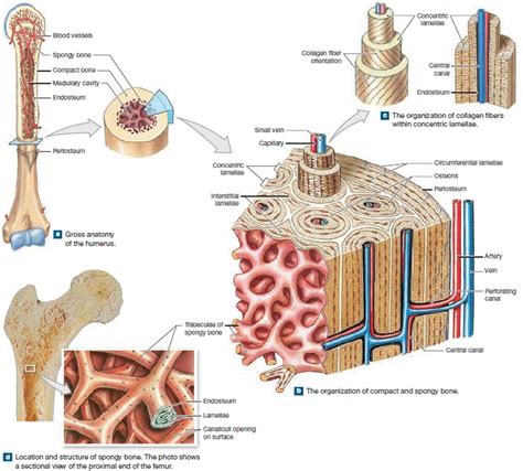
Table of Contents
The Structural Units of Mature Compact Bone are Called Osteons: A Deep Dive into Bone Histology
Mature compact bone, the dense outer layer of most bones, is a marvel of biological engineering. Its strength and resilience are crucial for supporting the body's weight and protecting vital organs. This strength is directly attributable to its highly organized microscopic structure. The fundamental structural units of mature compact bone are called osteons, also known as Haversian systems. Understanding osteons is key to comprehending how bone functions, repairs itself, and responds to stress. This article will delve into the intricate details of osteons, exploring their composition, arrangement, and significance in bone biology.
What are Osteons? A Detailed Look at the Haversian System
Osteons are cylindrical structures, roughly 4 millimeters in length and 0.2 millimeters in diameter, that run parallel to the long axis of long bones. Imagine them as tiny, intricately designed pillars holding up a magnificent structure. Each osteon is a complex assembly of several key components:
1. Central Canal (Haversian Canal): The Osteon's Core
At the heart of each osteon lies the central canal, also known as the Haversian canal. This canal acts as a vital conduit, running longitudinally through the osteon. It contains essential components for bone maintenance:
- Blood vessels: These provide oxygen and nutrients to the osteocytes, the bone cells residing within the osteon. This constant supply is crucial for bone metabolism and repair.
- Nerves: Nerves within the central canal transmit signals related to bone health and potential damage. They play a critical role in pain sensation and bone remodeling processes.
- Lymphatic vessels: These aid in removing waste products and maintaining fluid balance within the bone tissue.
The central canal is lined by a thin, delicate layer of connective tissue that supports the vascular and nervous components within.
2. Lamellae: Concentric Rings of Bone Matrix
Surrounding the central canal are concentric rings of lamellae. These lamellae are composed of collagen fibers and mineralized matrix, primarily hydroxyapatite crystals. The collagen fibers within each lamella are precisely arranged in a specific direction, and the direction changes slightly in adjacent lamellae. This alternating arrangement enhances the bone's overall strength and resistance to fracturing. The carefully orchestrated alignment of collagen fibers within each lamella and between adjacent lamellae plays a critical role in the bone's ability to withstand stress from multiple directions.
The concentric lamellae form the bulk of the osteon, showcasing nature's remarkable ability to create a strong, lightweight structure. The precise organization and arrangement of lamellae directly contributes to the overall strength and resilience of compact bone. The number of lamellae in an osteon can vary depending on the bone's location and the forces it routinely endures.
3. Lacunae: Homes for Osteocytes
Embedded within the lamellae are tiny spaces called lacunae. These lacunae are home to osteocytes, the mature bone cells. Each osteocyte occupies a single lacuna, residing within its own small compartment within the bone matrix. The osteocytes maintain and regulate the bone matrix surrounding them. Their long, slender processes, called canaliculi, extend through the matrix and connect with the processes of neighboring osteocytes.
This intricate network of interconnected canaliculi creates a communication pathway for the osteocytes. Nutrients and waste products are transported between the osteocytes via these canaliculi, effectively linking the cells to the central canal and the vascular supply. This interconnected network is essential for the survival and function of osteocytes within the osteon.
4. Canaliculi: Communication Pathways for Osteocytes
The canaliculi are a network of microscopic channels that radiate outward from the lacunae, connecting neighboring lacunae and eventually reaching the central canal. This complex network plays a crucial role in nutrient exchange and cellular communication within the osteon. The canaliculi facilitate the transport of nutrients and metabolic waste between the osteocytes and the blood vessels within the central canal. The intricate canalicular network ensures that even the most distant osteocytes receive the necessary nutrients and oxygen. This interconnectedness is crucial for maintaining the viability and proper function of the osteocytes and, consequently, the entire osteon.
Interstitial Lamellae: Remnants of Old Osteons
Between the intact osteons, you'll find interstitial lamellae. These are remnants of old osteons that have been partially or completely resorbed during bone remodeling. They are irregular fragments of lamellae, indicating the dynamic nature of bone tissue. The presence of interstitial lamellae showcases bone's constant state of remodeling and adaptation. Bone is not a static structure; it continuously remodels itself in response to mechanical stress and metabolic demands. Interstitial lamellae are a testament to this dynamic process, representing the remnants of previous osteon formations that have been partially resorbed and replaced.
Circumferential Lamellae: Encircling the Bone
Encircling the entire bone shaft are circumferential lamellae. These lamellae run parallel to the bone's surface, providing further structural support and reinforcing the overall strength of the compact bone. Similar to concentric lamellae, they consist of collagen fibers and mineralized matrix arranged in a highly organized fashion. Circumferential lamellae are located both internally (immediately beneath the periosteum) and externally (adjacent to the endosteum) contributing to the bone's overall structural integrity. The circumferential lamellae increase the bone's resistance to bending and twisting forces, ensuring its stability and load-bearing capacity.
The Significance of Osteons in Bone Function
The organized structure of osteons is crucial for several aspects of bone function:
- Strength and Support: The concentric arrangement of lamellae and the presence of mineralized matrix contribute to the exceptional strength and weight-bearing capacity of compact bone.
- Nutrient Supply: The central canal, with its blood vessels, ensures that all osteocytes receive adequate nutrients and oxygen, enabling their metabolic activities and facilitating bone maintenance.
- Waste Removal: The canaliculi and central canal provide pathways for removing metabolic waste products, maintaining a healthy environment for the osteocytes.
- Bone Remodeling: Osteons are involved in bone remodeling, a continuous process of bone resorption and formation that maintains bone integrity and adapts to changing mechanical stress. Osteoclasts resorb old bone, while osteoblasts lay down new bone matrix, constantly shaping and reshaping the osteons.
- Fracture Repair: During fracture repair, osteons play a crucial role in forming new bone tissue to bridge the gap between fracture fragments. The formation of new osteons is essential for the restoration of bone strength and integrity after a fracture.
The intricate structure of osteons ensures that bone is not just a strong and supportive tissue but also a highly dynamic and adaptive tissue capable of remodeling and repairing itself in response to various stimuli.
Clinical Significance of Osteon Structure
Understanding osteon structure is essential in various clinical settings:
- Osteoporosis: This condition, characterized by decreased bone density and increased fragility, involves alterations in osteon structure and remodeling. Osteoporosis often results in a reduced number of osteons and an altered arrangement of lamellae, making bones more susceptible to fracture.
- Bone Fractures: The structural properties of osteons influence fracture patterns and healing. The precise arrangement of collagen fibers in lamellae determines the bone's ability to withstand different types of mechanical stress.
- Bone Tumors: Tumors can alter the normal structure and organization of osteons, affecting the bone's mechanical properties and potentially leading to fractures.
- Paget's Disease: This bone disease involves disordered osteoclast and osteoblast activity, resulting in abnormal osteon formation and weakening of the bone structure.
Studying osteons helps researchers and clinicians understand bone diseases and develop effective treatments.
Conclusion: The Osteon – A Masterpiece of Biological Engineering
The osteon, the fundamental structural unit of mature compact bone, is a masterpiece of biological engineering. Its intricate design, featuring concentric lamellae, a central canal, lacunae, and canaliculi, combines strength, resilience, and efficient nutrient and waste transport. The understanding of osteon structure is paramount for comprehending bone's dynamic nature, its response to mechanical loading, and its capacity for repair and remodeling. Further research into osteon biology will undoubtedly lead to advancements in the treatment of bone diseases and the development of novel biomaterials for bone regeneration. The complexity and elegance of the osteon structure continue to inspire researchers and clinicians alike, highlighting the remarkable engineering principles embedded in the human body.
Latest Posts
Latest Posts
-
What Eats Seaweed In The Ocean
Mar 14, 2025
-
Cuanto Es 10 Cm En Pulgadas
Mar 14, 2025
-
Rewrite Imply With And And Or
Mar 14, 2025
-
What Is The Melting Temp Of Glass
Mar 14, 2025
-
The Transfer Of Energy As Electromagnetic Waves
Mar 14, 2025
Related Post
Thank you for visiting our website which covers about The Structural Units Of Mature Compact Bone Are Called . We hope the information provided has been useful to you. Feel free to contact us if you have any questions or need further assistance. See you next time and don't miss to bookmark.
