What Forms The Channels And Pumps In The Phospholipid Bilayer
Kalali
Mar 21, 2025 · 7 min read
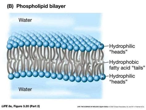
Table of Contents
What Forms the Channels and Pumps in the Phospholipid Bilayer?
The phospholipid bilayer, the fundamental structure of all cell membranes, isn't just a static barrier. Its remarkable functionality stems from a diverse array of embedded proteins that facilitate the selective transport of molecules across this otherwise impermeable membrane. These proteins form channels and pumps, sophisticated molecular machinery crucial for cellular life. Understanding their structure and function is key to comprehending cellular processes. This article delves into the intricate mechanisms that shape these vital components of the cell membrane.
The Phospholipid Bilayer: A Dynamic Barrier
Before diving into the protein structures, let's briefly revisit the phospholipid bilayer itself. This double layer of amphipathic phospholipids – molecules with both hydrophilic (water-loving) heads and hydrophobic (water-fearing) tails – arranges itself spontaneously in an aqueous environment. The hydrophilic heads face the extracellular and intracellular fluids, while the hydrophobic tails cluster together, creating a hydrophobic core that prevents the free passage of most polar molecules and ions. This fundamental structure provides the basis for selective permeability, a critical characteristic of cell membranes. The fluidity of the bilayer, influenced by factors like temperature and fatty acid composition, further contributes to its dynamic nature, allowing for the movement and interaction of membrane proteins.
Membrane Proteins: Architects of Transport
The remarkable functionality of the cell membrane is largely attributed to the diverse array of proteins embedded within the phospholipid bilayer. These membrane proteins can be broadly classified into integral and peripheral proteins. Integral membrane proteins are firmly embedded within the bilayer, often spanning the entire membrane (transmembrane proteins). Peripheral proteins, on the other hand, are loosely associated with the membrane, often interacting with the hydrophilic heads or integral proteins. Channels and pumps are primarily formed by integral membrane proteins.
Ion Channels: Selective Gates
Ion channels are transmembrane proteins that form aqueous pores allowing the passage of specific ions across the membrane. Their selectivity is crucial; they only permit the passage of certain ions, often excluding others, even if they are of similar size and charge. This selectivity is achieved through specific amino acid residues lining the pore, creating a molecular sieve that interacts with the ions based on their size, charge, and hydration shell.
Types of Ion Channels:
-
Voltage-gated channels: These channels open and close in response to changes in the membrane potential, a crucial mechanism in nerve impulse transmission and muscle contraction. The change in voltage alters the conformation of the channel protein, causing it to open or close the pore.
-
Ligand-gated channels: These channels are activated by the binding of a specific ligand, a molecule that binds to a receptor, to a receptor site on the channel protein. Neurotransmitters, for example, often act as ligands, binding to receptors on ligand-gated ion channels in postsynaptic neurons, triggering changes in membrane potential.
-
Mechanically-gated channels: These channels respond to mechanical forces applied to the membrane, such as stretch or pressure. They play a vital role in sensory perception, such as touch and hearing, where mechanical stimuli trigger the opening of channels.
Structure of Ion Channels:
The structure of ion channels varies greatly depending on the specific type of channel. Many are composed of multiple subunits, each contributing to the formation of the pore and its selectivity filter. The subunits often have transmembrane α-helices, which span the membrane and contribute to the structure of the pore. The arrangement of these helices and the amino acid residues within them dictate the size and charge selectivity of the channel. Detailed understanding of these structures often relies on techniques like X-ray crystallography and cryo-electron microscopy.
Pumps: Active Transporters
Unlike channels that facilitate passive transport (movement down a concentration gradient), pumps actively transport molecules against their concentration gradients, a process that requires energy, usually in the form of ATP hydrolysis. These pumps are also transmembrane proteins, but their structure and mechanism differ significantly from channels.
Types of Pumps:
-
P-type ATPases: These pumps are phosphorylated during the transport cycle, using ATP to drive the conformational change required for transport. The Na+/K+-ATPase, responsible for maintaining the sodium and potassium gradients across the cell membrane, is a prime example.
-
V-type ATPases: These pumps utilize ATP to pump protons (H+) across membranes, establishing a proton gradient often used to drive other transport processes. They are found in various organelles like lysosomes and vacuoles.
-
F-type ATPases (ATP synthases): While primarily functioning as ATP synthases, producing ATP from a proton gradient, they can also act as pumps under certain conditions, moving protons against their concentration gradient using the energy derived from ATP hydrolysis.
-
ABC transporters: These transporters use ATP to transport a wide variety of substrates, including ions, sugars, and lipids, across membranes. They have two ATP-binding domains that bind and hydrolyze ATP, driving the transport process.
Structure of Pumps:
Pumps typically have multiple transmembrane domains, often with specific binding sites for the transported molecule and ATP. The binding of ATP induces conformational changes in the pump protein, allowing it to bind the substrate, and then transport it across the membrane by altering the protein’s structure. The Na+/K+-ATPase, for instance, has multiple subunits and undergoes a complex series of conformational changes driven by ATP hydrolysis to move three sodium ions out of the cell and two potassium ions into the cell. The precise mechanisms and structures of different pumps have been elucidated through a combination of biochemical and structural biology techniques.
Regulation of Channels and Pumps: Maintaining Cellular Homeostasis
The activity of channels and pumps is precisely regulated to maintain cellular homeostasis. This regulation is crucial for various cellular processes, including nerve impulse transmission, muscle contraction, and nutrient uptake.
Mechanisms of Regulation:
-
Voltage-gating: As mentioned earlier, voltage changes can alter the conformation of channel proteins, controlling their opening and closing.
-
Ligand-gating: Binding of specific molecules (ligands) to receptor sites on channels or pumps can modulate their activity.
-
Phosphorylation: Covalent modification of channel or pump proteins by phosphorylation can alter their activity.
-
Second messenger signaling pathways: Intracellular signaling pathways triggered by extracellular stimuli can modulate the activity of channels and pumps.
-
Protein-protein interactions: Interactions with other membrane proteins or cytoplasmic proteins can regulate the activity of channels and pumps.
Diseases Associated with Dysfunction of Channels and Pumps
Malfunctions in ion channels and pumps are implicated in a wide range of diseases, highlighting the critical importance of their proper functioning.
Examples of Diseases:
-
Cystic fibrosis: Caused by mutations in the CFTR chloride channel, leading to impaired chloride ion transport and thick mucus buildup in the lungs and other organs.
-
Epilepsy: Certain forms of epilepsy are associated with mutations in ion channels, leading to abnormal neuronal excitability.
-
Long QT syndrome: Characterized by prolonged QT intervals in the electrocardiogram, associated with mutations in ion channels involved in cardiac repolarization.
-
Familial hypercholesterolemia: Caused by mutations in LDL receptors, affecting cholesterol uptake and leading to high cholesterol levels.
Future Directions: Unraveling the Complexity
The field of membrane transport continues to evolve. Advancements in structural biology, such as cryo-electron microscopy, are providing increasingly detailed views of channel and pump structures, revealing subtle conformational changes that govern their function. Computational modeling and simulations are further enhancing our understanding of transport mechanisms at the molecular level. This ongoing research is crucial not only for expanding our fundamental knowledge of cellular processes but also for developing novel therapeutic strategies for diseases associated with ion channel and pump dysfunction. As technology improves, we will continue to unveil the complex interplay between these essential components of the phospholipid bilayer.
Conclusion: The Intricate Dance of Life
The phospholipid bilayer, with its embedded channels and pumps, represents a remarkable example of biological sophistication. These protein structures, formed through intricate arrangements of amino acids and governed by complex regulatory mechanisms, are essential for life itself. Understanding their structure and function is fundamental to comprehending cellular processes, and ongoing research continues to unravel the intricate details of this vital dance of life. The precise interplay of these molecular machines dictates cell function, signaling, and overall homeostasis, and their dysfunction directly impacts human health. Continued study of channels and pumps holds the key to understanding the underlying mechanisms of a wide range of diseases, paving the way for innovative therapeutic interventions in the future.
Latest Posts
Latest Posts
-
How To Convert A Square Root Into A Decimal
Mar 22, 2025
-
4 To The Power Of 8
Mar 22, 2025
-
What Is 0 333 As A Fraction
Mar 22, 2025
-
What Is 39 Degrees Fahrenheit In Celsius
Mar 22, 2025
-
How Do You Turn A Mixed Number Into A Percent
Mar 22, 2025
Related Post
Thank you for visiting our website which covers about What Forms The Channels And Pumps In The Phospholipid Bilayer . We hope the information provided has been useful to you. Feel free to contact us if you have any questions or need further assistance. See you next time and don't miss to bookmark.
