What Is The Diaphragm Of A Microscope
Kalali
Mar 15, 2025 · 6 min read
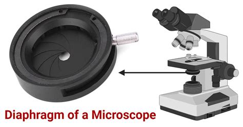
Table of Contents
What is the Diaphragm of a Microscope? A Comprehensive Guide
The microscope diaphragm, often overlooked, plays a crucial role in achieving high-quality images. Understanding its function is essential for any serious microscopy user, from students to seasoned researchers. This comprehensive guide delves deep into the world of microscope diaphragms, explaining their types, functions, and importance in achieving optimal image resolution and contrast.
Understanding the Role of the Diaphragm
The diaphragm in a microscope is an adjustable aperture located within the condenser. Its primary function is to control the amount of light that reaches the specimen. This seemingly simple function has a profound impact on image quality. By regulating light intensity and its distribution, the diaphragm influences:
-
Contrast: Proper diaphragm adjustment is crucial for achieving optimal contrast. Too much light can wash out details, while too little can result in a dark, unclear image. Finding the sweet spot maximizes the visibility of fine structures within the specimen.
-
Resolution: While resolution is primarily determined by the objective lens, the diaphragm plays a supporting role. Correct adjustment enhances the resolving power of the objective by optimizing the light cone entering the specimen.
-
Depth of Field: The diaphragm indirectly affects depth of field. A slightly closed diaphragm may improve depth of field, allowing more of the specimen to be in focus simultaneously. However, over-closing can negatively impact resolution and contrast.
Types of Microscope Diaphragms
Microscopes typically utilize one of the following types of diaphragms:
1. Iris Diaphragm: The Most Common Type
The iris diaphragm is the most prevalent type found in most modern microscopes. It consists of a series of overlapping metal plates that can be adjusted to precisely control the aperture size. A lever or a rotating wheel allows for smooth and gradual adjustments, offering fine control over light intensity. The iris diaphragm's versatility makes it ideal for a wide range of microscopy applications. Its ability to smoothly control light intensity allows users to optimize imaging parameters for various specimens and objectives.
Advantages of Iris Diaphragms:
- Precise Control: Offers precise control over light intensity.
- Versatility: Suitable for various microscopy techniques and specimens.
- Widely Available: Found in most modern microscopes.
2. Disc Diaphragm: A Simpler Alternative
The disc diaphragm is a simpler mechanism consisting of a rotating disc with several apertures of different sizes. Each aperture corresponds to a specific light intensity. The user simply rotates the disc to select the desired aperture. While less precise than the iris diaphragm, the disc diaphragm is often found in simpler, more affordable microscopes.
Advantages of Disc Diaphragms:
- Simplicity: Easy to use and understand.
- Cost-effectiveness: Typically found in budget-friendly microscopes.
3. Field Diaphragm: Controlling Illumination Evenness
While not strictly a part of the condenser diaphragm system, the field diaphragm deserves mention. Located at the base of the illuminator, the field diaphragm controls the size of the illuminated area on the specimen. Proper adjustment ensures even illumination and prevents stray light from impacting image quality. Adjusting the field diaphragm correctly ensures that only the area of the specimen under observation is illuminated, resulting in a sharper and better defined image.
How to Properly Adjust the Diaphragm
Proper diaphragm adjustment is crucial for optimal image quality. The process involves a careful balance to achieve the best compromise between brightness, contrast, and resolution. There's no single "correct" setting; the ideal position depends on the objective lens, the specimen, and the desired level of detail.
Here's a step-by-step guide:
-
Start with the diaphragm fully open: Begin with the diaphragm fully open to allow maximum light transmission.
-
Observe the image: Examine the image through the eyepiece. Note the brightness and contrast.
-
Gradually close the diaphragm: Slowly close the diaphragm, making small adjustments. Observe how the image changes. You'll likely see an increase in contrast but also a decrease in brightness.
-
Find the optimal setting: The optimal setting is typically found when the contrast is maximized without significantly sacrificing brightness or resolution. The optimal setting will showcase the specimen's detail as clearly as possible. Closing the diaphragm too much will result in diffraction effects, reducing the resolution.
-
Adjust for different objectives: The optimal diaphragm setting will vary depending on the magnification of the objective lens. Higher magnification objectives generally require a slightly more closed diaphragm.
-
Consider the specimen: Different specimens may require different diaphragm settings. Translucent specimens may need more light, while opaque specimens might benefit from a more closed setting.
-
Köhler Illumination: For more advanced microscopy, Köhler illumination should be used. This method ensures even illumination across the field of view. Köhler illumination requires careful adjustment of both the field diaphragm and the condenser diaphragm to achieve optimal results. This technique allows for the greatest image quality and ensures optimal use of the condenser diaphragm's capabilities.
The Importance of Diaphragm Adjustment in Different Microscopy Techniques
The diaphragm plays a significant role in various microscopy techniques, including:
-
Brightfield Microscopy: Proper diaphragm adjustment is crucial for maximizing contrast and resolution in brightfield microscopy.
-
Darkfield Microscopy: In darkfield microscopy, the diaphragm is used to create a hollow cone of light that illuminates the specimen from the sides, resulting in a bright specimen against a dark background. Precise diaphragm adjustment is essential for achieving optimal darkfield illumination.
-
Phase-Contrast Microscopy: Phase contrast uses the diaphragm to fine-tune the phase differences between the direct and diffracted light rays, enhancing contrast in transparent specimens.
-
Fluorescence Microscopy: While fluorescence microscopy relies on emitted light, the condenser diaphragm still plays a role in controlling stray light and improving image clarity.
-
Differential Interference Contrast (DIC) Microscopy: Similar to phase-contrast, DIC uses the diaphragm to manipulate light interference to enhance contrast.
Troubleshooting Common Diaphragm-Related Issues
If you're experiencing problems with your microscope images, the diaphragm might be the culprit. Here are some common issues and their solutions:
- Low contrast: Try closing the diaphragm slightly.
- Poor resolution: Ensure the diaphragm is not closed too much. Opening it slightly might improve resolution.
- Uneven illumination: Check the field diaphragm for proper adjustment. Also, ensure the condenser is correctly aligned.
- Diffraction artifacts: Opening the diaphragm slightly can resolve diffraction artifacts caused by over-closing.
Conclusion: Mastering the Art of Diaphragm Adjustment
The microscope diaphragm is a deceptively simple yet powerful tool that significantly influences image quality. Understanding its function and mastering its adjustment is essential for achieving optimal results in microscopy. By carefully adjusting the diaphragm, you'll unlock the full potential of your microscope, revealing the intricate details of your specimens with clarity and precision. Proper diaphragm usage, alongside Köhler illumination techniques when applicable, is paramount to obtaining high-quality images, regardless of the type of microscopy employed. Regular practice and keen observation will allow you to become proficient in its use, ultimately leading to more impactful microscopy experiences. The seemingly simple act of adjusting this often-overlooked component is a crucial step toward achieving truly exceptional microscopy results.
Latest Posts
Latest Posts
-
What Is 0 2 As A Percentage
Mar 15, 2025
-
How Many Combinations Of Phone Numbers Are There
Mar 15, 2025
-
Common Multiple Of 3 4 5
Mar 15, 2025
-
What Is 112 F In Celsius
Mar 15, 2025
-
Cuanto Es 35 Pulgadas En Metros
Mar 15, 2025
Related Post
Thank you for visiting our website which covers about What Is The Diaphragm Of A Microscope . We hope the information provided has been useful to you. Feel free to contact us if you have any questions or need further assistance. See you next time and don't miss to bookmark.
