When Beginning To Focus Use The Scanning Objective Lens
Kalali
Mar 26, 2025 · 6 min read
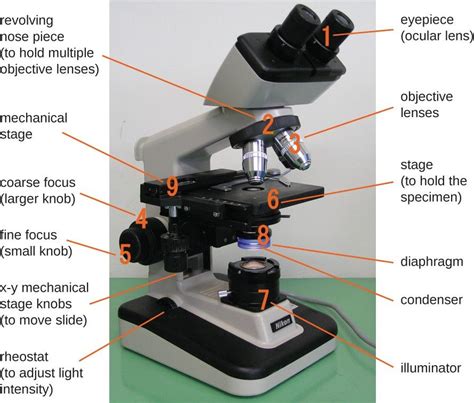
Table of Contents
When Beginning to Focus, Use the Scanning Objective Lens: A Comprehensive Guide to Microscopy
Microscopy, the art of visualizing the incredibly small, is a cornerstone of scientific discovery across numerous fields. From biology and medicine to materials science and engineering, understanding how to effectively use a microscope is paramount. A crucial, often overlooked, step in this process is starting your observation with the scanning objective lens. This seemingly simple act dramatically improves your workflow, protects your equipment, and enhances the accuracy of your observations. This comprehensive guide will delve into the reasons why you should always begin with the scanning objective lens, how to do it correctly, and the benefits it provides.
Why Start with the Scanning Objective Lens?
The scanning objective lens, typically the 4x lens, is the lowest magnification lens on most compound light microscopes. Its primary role isn't just about low magnification; it's about setting the stage for successful high-magnification viewing. Ignoring this crucial first step can lead to frustration, damaged equipment, and inaccurate results. Here's a breakdown of the key reasons:
1. Finding the Specimen: The Initial Search
Imagine trying to find a specific grain of sand on a vast beach without a map. That's essentially what you're doing when you jump straight to high magnification without first locating your specimen with the scanning lens. The field of view at higher magnifications is drastically smaller, making it incredibly difficult, if not impossible, to find your target. The 4x lens provides a wide field of view, allowing you to quickly survey the slide and locate the area of interest.
2. Preventing Slide Damage: Avoiding Collision
High-power objective lenses have a very short working distance – the distance between the lens and the slide. If you attempt to focus at high magnification without first locating your specimen at low magnification, there's a significant risk of crashing the objective lens into the slide. This can damage both the lens and the slide, potentially resulting in costly repairs or replacement. Starting with the scanning lens provides ample working distance, minimizing this risk.
3. Efficient and Effective Focusing: A Gradual Approach
Focusing at high magnification is a delicate process. Small adjustments can mean the difference between a clear image and a blurry mess. Starting with the scanning lens allows for coarse focusing, quickly bringing the specimen into approximate focus. You can then systematically increase magnification, making fine adjustments at each stage for optimal clarity. This graduated approach is far more efficient and effective than attempting to focus directly at high magnification.
4. Improved Image Quality: Sharpness and Clarity
While the scanning objective lens offers the lowest magnification, its broad field of view and large working distance contribute to a generally sharper image. This is because there are fewer optical elements to potentially distort the image. You can avoid out-of-focus areas and create a much more pleasing base view from which to build higher magnifications. Trying to start with high power magnifications without having established a clear and focused base layer at 4x, often leads to very poor image quality that may be impossible to remedy.
5. Prolonging Microscope Lifespan: Preventing Wear and Tear
Repeatedly crashing high-power objective lenses into slides places considerable stress on the delicate optical components. This wear and tear can significantly shorten the lifespan of your microscope, leading to costly repairs or premature replacement. Starting with the scanning lens helps prevent this damage, contributing to a longer operational life of your valuable equipment.
The Step-by-Step Process: Mastering the Technique
Now that we've established the importance of starting with the scanning objective lens, let's examine the correct procedure. This should be part of every microscopy session:
1. Prepare Your Slide: Ensure your slide is clean, properly mounted, and securely positioned on the microscope stage.
2. Select the Scanning Objective Lens (4x): Rotate the objective turret until the 4x lens clicks into place. This is usually the shortest objective lens.
3. Coarse Focusing: Use the coarse focus knob (typically the larger knob) to adjust the height of the stage, bringing the specimen into approximate focus. Move the stage slowly and carefully to avoid collisions.
4. Fine Focusing: Once the specimen is approximately in focus, use the fine focus knob (typically the smaller knob) for precise adjustments, achieving a sharp, clear image.
5. Center Your Specimen: Once focused, center the area of interest within the field of view. This ensures it remains visible as you increase magnification.
6. Increase Magnification Gradually: Carefully rotate the objective turret to the next higher magnification lens (e.g., 10x). Only minimal fine focus adjustments should be necessary at this stage.
7. Repeat the Process: Continue increasing magnification in small steps (e.g., 10x to 40x) with fine focusing at each stage. Never go directly from the 4x to the 100x oil immersion lens! Remember, at each stage, make sure the specimen remains centered and clear.
8. Oil Immersion (If Applicable): For the highest magnification (typically 100x), apply a small drop of immersion oil to the slide before carefully rotating the 100x objective lens into position. Remove oil and clean the lens immediately after use.
9. Documentation: Once you've achieved the desired magnification and clarity, document your findings through drawings, photographs, or digital imaging.
Beyond the Basics: Advanced Techniques and Considerations
While the core principle of starting with the scanning lens remains constant, several advanced techniques and considerations can further enhance your microscopy experience:
Köhler Illumination: Optimizing Image Quality
Köhler illumination is a method of adjusting the microscope's lighting to achieve optimal evenness and clarity. Proper Köhler illumination is crucial for obtaining high-quality images at all magnifications. This process should be performed before you begin focusing.
Working Distance Awareness: Maintaining Safety
Always be mindful of the working distance of each objective lens. Never force the lens closer to the slide than the lens design permits, particularly at higher magnifications. It is important to be familiar with the working distances of all your objective lenses.
Slide Preparation: Ensuring Clarity
The quality of your slide preparation directly impacts the clarity of your microscopic images. Using proper staining techniques and ensuring your specimen is thin and evenly distributed across the slide are vital to successful microscopy.
Microscope Maintenance: Protecting your Investment
Regular cleaning and maintenance of your microscope are crucial to ensuring its long-term performance and accuracy. Clean lenses gently with lens cleaning paper and appropriate solvents. Avoid harsh chemicals that could damage the lens coatings.
Troubleshooting: Addressing Common Issues
Despite following proper procedures, you may encounter issues like blurry images or inability to focus at high magnification. Carefully check for issues such as incorrect lighting, dirty lenses, improper slide preparation, or mechanical problems with the microscope. If the problem is persistent, consult your microscope's manual or seek assistance from a microscopy expert.
Conclusion: The Foundation of Successful Microscopy
Starting your microscopy observations with the scanning objective lens is not merely a suggestion; it’s a fundamental best practice. This simple yet crucial step significantly improves your efficiency, protects your equipment, and ultimately leads to higher-quality images and more accurate scientific observations. By mastering this technique and incorporating the advanced tips discussed, you will greatly enhance your proficiency in microscopy, regardless of your field of study or level of experience. Remember that consistent practice and attention to detail are key to becoming a successful microscopist. The rewards are well worth the effort: the ability to visualize the hidden wonders of the microscopic world, unlocking new discoveries and deepening our understanding of the universe at its smallest scale.
Latest Posts
Latest Posts
-
What Is 18 25 As A Percentage
Mar 29, 2025
-
29 C Is What In Fahrenheit
Mar 29, 2025
-
Cuanto Es 52 Fahrenheit En Centigrados
Mar 29, 2025
-
What Base Is Found On Rna But Not On Dna
Mar 29, 2025
-
What Is 5 5 As A Decimal
Mar 29, 2025
Related Post
Thank you for visiting our website which covers about When Beginning To Focus Use The Scanning Objective Lens . We hope the information provided has been useful to you. Feel free to contact us if you have any questions or need further assistance. See you next time and don't miss to bookmark.
