Why Does Dna Move To The Positive Electrode
Kalali
Mar 16, 2025 · 6 min read
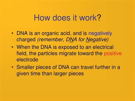
Table of Contents
Why Does DNA Move to the Positive Electrode? Understanding Electrophoresis
DNA, the fundamental blueprint of life, carries the genetic instructions for all living organisms. While seemingly static within a cell's nucleus, DNA's behavior under certain conditions reveals fascinating aspects of its molecular structure and properties. One such phenomenon is its migration towards the positive electrode during electrophoresis, a technique widely used in molecular biology and genetics. This article delves deep into the reasons behind this movement, exploring the underlying principles of electrophoresis and the factors influencing DNA's electrophoretic mobility.
Understanding Electrophoresis: The Driving Force Behind DNA Movement
Electrophoresis is a powerful laboratory technique that separates charged molecules based on their size and charge. This separation occurs when the molecules are placed in an electric field, causing them to migrate towards the electrode with the opposite charge. In the context of DNA, this means that negatively charged DNA fragments move towards the positive electrode (anode). But why is DNA negatively charged in the first place?
The Phosphate Backbone: The Source of Negative Charge
The key to understanding DNA's movement lies in its chemical structure. DNA is a double-stranded helix composed of nucleotides. Each nucleotide consists of a deoxyribose sugar, a phosphate group, and a nitrogenous base (adenine, guanine, cytosine, or thymine). Crucially, it's the phosphate group that imparts the negative charge to the DNA molecule.
The phosphate group possesses a negatively charged oxygen atom. These negatively charged phosphate groups form the backbone of the DNA molecule, giving the entire DNA strand a significant overall negative charge. This negative charge is independent of the DNA sequence and consistently present in every DNA molecule.
The Electrophoretic Field: Guiding the Movement of DNA
The electric field applied during electrophoresis provides the driving force for DNA migration. The electric field is created by applying a voltage across a gel matrix, typically agarose or polyacrylamide. This creates a potential difference, forcing the negatively charged DNA fragments to move towards the positive electrode. The strength of the electric field, expressed in volts per centimeter (V/cm), influences the speed at which the DNA migrates. A stronger field leads to faster migration.
The Gel Matrix: A Sieve for DNA Separation
The gel matrix isn't simply a passive medium; it plays a crucial role in separating DNA fragments based on size. The gel acts as a molecular sieve, hindering the movement of larger DNA fragments more than smaller ones. This is because larger DNA molecules have a greater difficulty navigating the pores within the gel matrix. Smaller DNA fragments, with their greater maneuverability, travel faster through the gel.
Agarose and Polyacrylamide Gels: Different Sieving Properties
Agarose gels, commonly used for separating larger DNA fragments (several hundred base pairs to tens of kilobases), have larger pore sizes. Polyacrylamide gels, on the other hand, possess smaller pores and are typically used to separate smaller DNA fragments (tens to thousands of base pairs). The choice of gel depends on the size range of the DNA fragments being analyzed.
Factors Influencing DNA Mobility in Electrophoresis
Several factors can influence the electrophoretic mobility of DNA fragments, affecting their migration speed and separation efficiency:
1. DNA Size: A Major Determinant of Mobility
As mentioned earlier, the size of the DNA fragment is a primary determinant of its mobility. Smaller fragments migrate faster than larger fragments due to their increased ability to navigate the gel matrix's pores. This size-based separation is the foundation of DNA fingerprinting and other molecular biology techniques.
2. Gel Concentration: Fine-Tuning Separation
The concentration of the gel matrix directly influences the pore size. Higher gel concentrations result in smaller pores, reducing the mobility of all DNA fragments but increasing the resolution of separation between fragments of similar sizes. Conversely, lower gel concentrations create larger pores, increasing mobility but potentially reducing resolution.
3. Voltage: Speed Control
The applied voltage affects the speed of DNA migration. Higher voltages lead to faster migration but can also generate heat, which can damage the DNA or distort the gel. Optimizing the voltage is crucial for efficient separation without compromising the integrity of the sample.
4. Buffer Composition: Maintaining Optimal pH and Conductivity
The buffer solution used in electrophoresis plays a critical role in maintaining the pH and ionic strength of the system. The buffer helps to ensure that the DNA remains negatively charged and conducts the electric current efficiently. Different buffers have different properties, and the selection depends on the specific application.
5. DNA Conformation: Supercoiling and Relaxation
The conformation of the DNA molecule can influence its mobility. Supercoiled DNA, which is tightly twisted, migrates slower than relaxed circular DNA or linear DNA of the same size. This difference in mobility can be exploited to study DNA topology and the action of enzymes that affect DNA structure.
6. Temperature: Controlling Diffusion and Mobility
Temperature can affect DNA mobility by altering the viscosity of the buffer and the gel matrix. Increased temperature can lead to increased diffusion, reducing the sharpness of DNA bands. Maintaining a consistent temperature is crucial for optimal resolution.
Applications of DNA Electrophoresis: A Wide Range of Uses
DNA electrophoresis is an indispensable technique in various molecular biology and genetic applications:
1. DNA Fingerprinting: Forensic Science and Paternity Testing
DNA fingerprinting, based on the analysis of variable number tandem repeats (VNTRs), relies on electrophoresis to separate DNA fragments of different sizes, generating unique patterns for individual identification. This has profound implications in forensic science, paternity testing, and other areas of human identification.
2. Restriction Fragment Length Polymorphism (RFLP) Analysis: Genetic Mapping and Disease Diagnosis
RFLP analysis utilizes restriction enzymes to cut DNA at specific sequences, generating fragments of different sizes. Electrophoresis separates these fragments, allowing for the detection of variations in DNA sequences linked to genetic diseases or other traits.
3. Polymerase Chain Reaction (PCR) Product Analysis: Amplicon Size Determination
PCR amplifies specific DNA sequences, creating numerous copies of a target fragment. Electrophoresis is crucial for determining the size of the PCR product, verifying the success of the amplification reaction and confirming the presence of the target DNA sequence.
4. Gene Cloning and Sequencing: Analyzing DNA Fragments
Electrophoresis plays a role in several steps of gene cloning and DNA sequencing. The technique is essential for verifying the size of cloned DNA fragments and assessing the quality of sequencing reactions.
5. DNA Quantification: Estimating DNA Concentration
Electrophoresis can provide a rough estimate of DNA concentration by comparing the intensity of DNA bands to standards. While not as accurate as spectrophotometric methods, this technique can be useful for quick assessments.
Conclusion: Understanding the Electrophoretic Journey of DNA
The movement of DNA towards the positive electrode during electrophoresis is a direct consequence of the negative charge imparted by the phosphate backbone. This seemingly simple phenomenon underpins a powerful technique with wide-ranging applications in molecular biology and genetics. Understanding the principles of electrophoresis, including the role of the electric field, gel matrix, and various influencing factors, is crucial for interpreting experimental results and exploiting the full potential of this fundamental laboratory technique. As technology continues to advance, electrophoresis remains an integral part of the ever-evolving landscape of molecular biology research, promising further breakthroughs in our understanding of life's intricate code.
Latest Posts
Latest Posts
-
24 Feet Is How Many Meters
Mar 17, 2025
-
The Property Of Volume Is A Measure Of
Mar 17, 2025
-
Distance From Earth To The Sun In Light Years
Mar 17, 2025
-
How Are Photosynthesis And Cellular Respiration Interrelated
Mar 17, 2025
-
How Much Is 50 Of 8
Mar 17, 2025
Related Post
Thank you for visiting our website which covers about Why Does Dna Move To The Positive Electrode . We hope the information provided has been useful to you. Feel free to contact us if you have any questions or need further assistance. See you next time and don't miss to bookmark.
