The Shaft Of The Long Bone Is Called
Kalali
Mar 31, 2025 · 6 min read
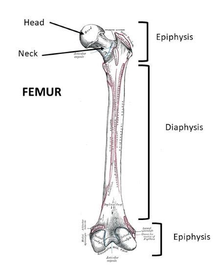
Table of Contents
The Shaft of the Long Bone is Called the Diaphysis: A Deep Dive into Long Bone Anatomy
The human skeletal system, a marvel of biological engineering, provides structure, support, and protection for our bodies. Central to this system are the long bones, crucial for locomotion, manipulation, and overall skeletal integrity. Understanding the anatomy of these bones is fundamental to appreciating their function and the impact of various conditions affecting them. This article will delve into the detailed anatomy of the long bone, focusing specifically on the shaft, which is called the diaphysis.
What is a Long Bone?
Before we explore the diaphysis, let's define what constitutes a long bone. Long bones are characterized by their elongated shape, significantly longer than they are wide. They are primarily found in the appendages – the arms and legs – and contribute significantly to height and limb movement. Examples include the femur (thigh bone), tibia (shin bone), fibula (calf bone), humerus (upper arm bone), radius (forearm bone), and ulna (forearm bone). While the term "long" might seem straightforward, the classification is based on shape rather than absolute length; some long bones in smaller animals are surprisingly short.
The Diaphysis: The Core of the Long Bone
The diaphysis, or shaft, forms the main, elongated portion of the long bone. It’s the strong, cylindrical part responsible for resisting bending and torsional forces during weight-bearing activities and movement. The diaphysis's unique structure contributes significantly to its strength and resilience.
Composition of the Diaphysis:
The diaphysis is primarily composed of:
-
Compact Bone: This dense, hard outer layer provides the structural strength and rigidity to withstand significant stress. The compact bone is organized in concentric lamellae (rings) around Haversian canals, containing blood vessels and nerves. This organized structure, known as an osteon, is highly efficient at distributing stress evenly across the bone.
-
Medullary Cavity: Within the diaphysis lies the medullary cavity, a hollow cylindrical space. In adults, this cavity is filled with yellow bone marrow, primarily composed of adipose (fat) tissue. Yellow bone marrow plays a role in energy storage and hematopoiesis (blood cell production) under certain conditions, though its primary role in adults is fat storage. In infants and young children, the medullary cavity is filled with red bone marrow, which is actively involved in hematopoiesis.
The Periosteum: A Protective Covering
The diaphysis is covered by a fibrous connective tissue membrane called the periosteum. This membrane plays several crucial roles:
-
Bone Growth and Repair: The periosteum contains osteoblasts, cells responsible for bone formation. It is essential for bone growth in width (appositional growth) and for fracture repair.
-
Nutrient Supply: The periosteum is richly supplied with blood vessels that penetrate the bone through perforating canals (Volkmann's canals), nourishing the underlying bone tissue. These canals connect to the Haversian canals within the compact bone.
-
Attachment Point for Muscles and Tendons: The periosteum provides attachment points for tendons and ligaments, allowing muscles to exert forces on the bone and facilitating movement.
The Metaphysis: The Transition Zone
At either end of the diaphysis, the bone transitions into the metaphysis. This region is characterized by a widening of the bone, connecting the diaphysis to the epiphyses (the ends of the long bone).
The Growth Plate: A Crucial Area for Longitudinal Bone Growth
The metaphysis contains the epiphyseal plate, also known as the growth plate. This is a layer of hyaline cartilage responsible for longitudinal bone growth during childhood and adolescence. The epiphyseal plate consists of zones of actively proliferating cartilage cells, which eventually ossify (become bone) as they mature. Once growth ceases, typically in late adolescence or early adulthood, the epiphyseal plate closes, leaving behind the epiphyseal line, a remnant of the former growth plate.
The Epiphyses: The Ends of the Long Bone
The epiphyses are the ends of the long bone. They are covered with articular cartilage, a specialized type of cartilage that reduces friction at the joints. The epiphyses also contain spongy bone, also known as cancellous bone, a lighter, less dense bone tissue than the compact bone of the diaphysis. Spongy bone contains trabeculae, a network of interconnected bony struts, providing structural support while minimizing weight. The spaces within spongy bone are filled with red bone marrow, which is actively involved in hematopoiesis throughout life.
Clinical Significance of Diaphyseal Anatomy
Understanding the diaphysis's anatomy is crucial in various clinical scenarios:
-
Fractures: Diaphyseal fractures are common, often resulting from high-impact trauma. The location and type of fracture significantly influence treatment strategies, which may range from casting to surgical intervention.
-
Bone Infections (Osteomyelitis): The diaphysis can be affected by osteomyelitis, a severe bone infection, often requiring aggressive antibiotic treatment and potentially surgical debridement (removal of infected tissue).
-
Bone Tumors: Primary bone tumors can originate in the diaphysis, requiring diagnosis through imaging techniques and potential surgical resection or other treatment modalities.
-
Bone Marrow Aspirations and Biopsies: The medullary cavity, accessible through the diaphysis, provides a site for bone marrow aspirations and biopsies, crucial for diagnosing hematologic diseases.
-
Intramedullary Nailing: Diaphyseal fractures are frequently treated with intramedullary nailing, a surgical procedure involving the insertion of a rod into the medullary cavity to stabilize the fracture.
The Diaphysis in Relation to Other Long Bone Components
The diaphysis works in concert with the metaphyses and epiphyses to form a functional unit. The diaphysis provides the primary structural support and resistance to bending forces, while the metaphyses allow for longitudinal growth and act as transition zones. The epiphyses contribute to joint articulation and provide additional support. This integrated design allows the long bone to efficiently perform its role in movement and weight-bearing.
Conclusion: A Functional Masterpiece
The diaphysis, or shaft, is not just a simple component of the long bone; it is a functionally significant structure intricately designed to withstand stress, provide support, and house the medullary cavity. Its compact bone composition, rich vascular supply via the periosteum and Haversian canals, and internal medullary cavity contribute to its strength and overall function. Understanding the diaphysis's anatomy is paramount for clinicians, researchers, and anyone interested in the intricacies of the human skeletal system and its remarkable capacity for movement, support, and adaptation. The diaphysis, in conjunction with the other elements of the long bone, is a testament to the elegance and efficiency of biological design. Further research into the diaphysis and long bone development continues to reveal fascinating insights into growth, repair mechanisms, and the overall resilience of our skeletal structure. As our understanding evolves, the focus on optimizing bone health and treating bone-related disorders will continue to benefit from this knowledge.
Latest Posts
Latest Posts
-
How To Find Length Of Chord
Apr 01, 2025
-
What Is The Speed Of Sound Mph
Apr 01, 2025
-
Convert 1 1 2 Inches To Millimeters
Apr 01, 2025
-
41 Degrees Celsius Converted To Fahrenheit
Apr 01, 2025
-
Is 0 0000008 J A Little Kinetic Enegy
Apr 01, 2025
Related Post
Thank you for visiting our website which covers about The Shaft Of The Long Bone Is Called . We hope the information provided has been useful to you. Feel free to contact us if you have any questions or need further assistance. See you next time and don't miss to bookmark.
